
Rotoscoliosis is a specific type of scoliosis that involves a significant rotational component in addition to the typical lateral...

Scoliosis, a condition characterized by an abnormal curvature of the spine, is particularly concerning when diagnosed in young children...
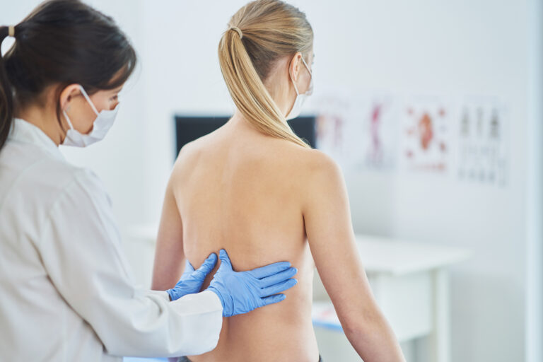
Degenerative scoliosis, a condition marked by the abnormal curvature of the spine, is often perceived as a debilitating ailment....
Scoliosis, a lateral curvature of the spine, is a condition that affects individuals across their lifespan, presenting unique challenges...

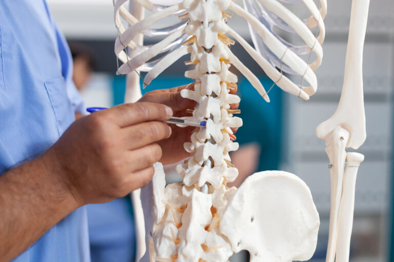
The human spine is a complex and vital structure that provides support and flexibility to the body. However, various...

Scoliosis, a condition characterized by an abnormal curvature of the spine, often prompts individuals to explore various forms of...